
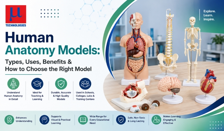
Human Anatomy Models: Types, Uses, Benefits & How to Choose the Right Model
Human anatomy models have quietly become one of the most important tools in modern medical, nursing, dental, veterinary, and biology education. Long before anyone could run a virtual dissection on a tablet, teachers needed a way to show students what the inside of the human body actually looks like — without a cadaver on hand every time. That need is exactly what gave rise to the anatomy model industry, and it's why the market for these models keeps expanding today.
Whether you're a school buying your first skeleton, a nursing college outfitting a simulation lab, a clinic that wants better tools for patient counseling, or a hobbyist curious about the science behind these teaching aids, this guide walks through everything worth knowing: what anatomy models are, the different types available, what they're used for, the evidence behind their benefits, and a practical framework for choosing the right one.
What Is a Human Anatomy Model?
A human anatomy model is a three-dimensional physical (or digital) representation of the human body or a specific part of it, built to teach anatomy and physiology without needing a real cadaver. These models range from a simple plastic skull to a full-body, multi-part torso with removable organs, and they're used across medical schools, nursing programs, dental colleges, veterinary schools, hospitals, physiotherapy clinics, and even secondary-school biology classrooms
A Quick History
Anatomy models are not a new idea — they're centuries old, and their history explains a lot about why they exist at all.
- Between the 16th and 19th centuries, the most common anatomical models were made of wax, partly because cadavers were difficult to obtain due to religious and legal restrictions on dissection.
- In the early 19th century, French medical student Louis Thomas Jérôme Auzoux grew frustrated that cadavers decomposed too quickly to study properly, while wax models were fragile and expensive. After visiting a papier-mâché workshop, he developed a durable paper-pulp technique that let models be taken apart piece by piece and reassembled — essentially inventing the first mass-producible "dissectible" anatomy model.
- Auzoux presented his first model to the Paris Academy of Medicine in 1822, and by 1827 had opened a dedicated factory in Normandy to keep up with international demand, eventually producing human, veterinary, and botanical models that were shipped as far as the Ottoman Empire.
- By the late 19th century, plaster of Paris became a popular, more affordable alternative, and through the 20th century the industry gradually shifted to plastic, rubber, and synthetic resins — the materials still dominant today.
That two-century-old problem — cadavers are scarce, expensive, and hard to preserve — is still the core reason anatomy models exist, and it's still driving the market today.
Why Anatomy Models Still Matter: The Cadaver Problem
Cadaveric dissection is widely regarded by anatomists as the richest way to learn anatomy, but it comes with real, well-documented constraints:
- Cost. One published cost breakdown from a U.S. medical school cadaver review session put the price of a single cadaver at roughly $1,900, with lab fees, supplies, dissection fees, and anatomist supervision pushing the total cost for a two-year teaching cycle to about $13,650.
- Supply shortages. In Karnataka, India, government medical colleges received fewer than 700 cadavers over three years against a requirement of roughly 900, even though national guidelines call for one cadaver per 10 MBBS students. Similar shortages have been documented at the University of Nairobi, where the cadaver-to-student ratio worsened from 1:6 to 1:9 despite growing enrollment .
- COVID-era disruption. The pandemic intensified cadaver shortages in the U.S. and accelerated many schools' shift toward models and virtual anatomy tools.
- Ethical and cultural limits. In some countries, including Taiwan, cultural norms around respect for the deceased significantly restrict the number of donated bodies available for teaching, pushing institutions toward plastic models and prosection.
Anatomy models solve the reusability problem that cadavers can't: a single well-made model can be disassembled and reassembled by hundreds of students over many years without decomposing, without the need for cold storage or preservation chemicals, and without the ethical and legal complications of handling human remains.
Read More: Best Human Skeleton Model Supplier in India
Types of Human Anatomy Models
Anatomy models are generally grouped by scope and function. Here are the main categories you'll come across when shopping for one.
1. Whole-Body / Full Skeleton Models
These display the complete human skeletal structure, sometimes with painted muscle origins and insertions, and are available in disarticulated, half, or full life-size formats. Detailed versions can show 600+ hand-numbered anatomical landmarks.
2. Torso Models
A torso model — often with a dissectible head — shows the position of internal organs (heart, lungs, liver, stomach, intestines, kidneys) within the trunk, and typically allows individual organs to be lifted out for close study.
3. Regional / Organ-Specific Models
These focus on one organ or body region in detail — brain, heart, eye, ear, kidney, spine, larynx, or joints — and are common where a course needs deep detail on a single system rather than a broad overview.
4. Detachable / Dissectible Models
Following the same logic as Auzoux's original 19th-century design, these models come apart into numbered pieces so learners can "dissect" and reassemble them repeatedly, simulating the exploratory process of real dissection without any of the risks.
5. Cross-Section / Transparent Models
Some models are built with transparent shells or sliced cross-sections so learners can see internal structure and spatial relationships from the outside in — useful for teaching how organs sit relative to one another.
6. Procedural / Skills-Training Models
Not every model is meant purely for visual reference. Suture pads, venipuncture and injection training arms, and acupuncture models exist specifically so students, nurses, and medical assistants can practice hands-on clinical skills before working on a real patient .
7. Digital and Virtual Anatomy Models
Digital anatomical models are built by scanning and rendering thin slices of scanned human bodies, and today extend to full 3D, VR, and mixed-reality anatomy platforms that let students rotate, layer, and "walk through" anatomical structures on a screen or headset.
8. 3D-Printed Anatomical Models
Increasingly, physical anatomy models are produced with additive manufacturing (3D printing) rather than injection-molded plastic, allowing for patient-specific replicas built directly from a real patient's CT or MRI scan — used heavily in surgical planning as well as education.
What Materials Are Anatomy Models Made From?
Most modern models are built from plastic, PVC/vinyl, rubber, or silicone, chosen for their durability, realistic texture, and ease of cleaning and maintenance. Silicone tends to be used where a soft, tissue-like feel matters most (such as suture pads or injection trainers), because it holds up to repeated puncturing better than more rigid plastics, though even silicone still can't fully replicate the exact tactile feel of real human tissue. High-end organ and skeletal models are typically hand-painted and numbered to identify structures precisely, which is part of why detailed models cost more than basic ones.
Where Human Anatomy Models Are Used
Anatomy models show up in more settings than most people expect:
- Medical, nursing, dental, and allied-health education — the largest and most obvious use case, spanning classroom lectures, simulation labs, and skills-training programs.
- Veterinary schools — anatomical models aren't limited to humans; canine, feline, and other animal models serve veterinary anatomy education the same way human models serve medical schools.
- Clinical patient communication — GP practices and physiotherapists frequently keep anatomy models on hand to visually explain a diagnosis, treatment, or upcoming procedure to a patient in a way words alone can't do as clearly .
- K-12 and general science education — simplified, smaller-scale models are widely used in school biology classes to introduce children to human anatomy.
- Surgical planning — patient-specific 3D-printed models built from a patient's own imaging data are increasingly used by surgeons to rehearse complex procedures before operating.
- Research and simulation centers — academic institutes represented the largest end-user segment of the broader medical simulation market in 2025, according to industry analysis, with hospitals expected to be the fastest-growing segment going forward.
Do Anatomy Models Actually Improve Learning? What the Research Shows
This is the part most marketing pages skip, but it's the most important for a buyer to know — and there's real published research behind it.
A 2020 systematic review and meta-analysis in BMC Medical Education pooled data from multiple studies comparing 3D-printed anatomy models to conventional teaching methods. It found that students using 3D-printed models scored meaningfully better on post-training tests in neuroanatomy, cardiac anatomy, and abdominal anatomy than students taught with conventional methods, and also performed better than students using standard 2D materials. The same review found students completed anatomy-related tasks faster with 3D models, and reported higher satisfaction with them compared to conventional teaching in most of the studies reviewed .
More recent research backs this up from a different angle. A 2025 study published in Frontiers in Medicine tested 169 medical students split into groups using traditional blended learning versus groups that incorporated continuous or phased 3D anatomy model integration. The group using continuous 3D-model integration scored significantly higher than the traditional group at multiple assessment stages .
The underlying reason researchers give for this is fairly intuitive: understanding anatomy requires strong 3D spatial reasoning, and static 2D textbook images and slides struggle to convey the depth, layering, and spatial relationships that a physical or 3D digital model can show directly. Handling, rotating, and disassembling a model gives learners a kind of hands-on engagement that a flat image simply can't replicate .
That said, the research is nuanced, not one-sided. Direct cadaveric dissection still lets students feel real tissue texture and appreciate anatomical variation in a way no model fully replicates, and several anatomists argue dissection remains the gold standard where it's available. The realistic takeaway: anatomy models are not necessarily a full replacement for cadaveric dissection where dissection is available, but they are a well-evidenced, cost-effective, and increasingly necessary supplement — and, where cadavers simply aren't accessible, a proven substitute.
Practical Benefits of Anatomy Models (Beyond Test Scores)
Pulling from the sources above, the concrete, sourced benefits are:
- Reusability — a single model can be used by generations of students, unlike a cadaver, which is typically used for one academic cycle and then must be scientifically disposed of.
- Lower long-term cost relative to sourcing, preserving, and disposing of cadavers, especially once you factor in lab fees, preservation chemicals, and specialist staff time.
- No ethical or religious barriers to access, which matters enormously in regions where cultural norms restrict body donation.
- Improved test performance and satisfaction, as shown in the meta-analysis and 2025 study cited above.
- Better patient communication in clinical settings, where a model helps a non-medical patient visually understand a diagnosis or procedure far more easily than a verbal explanation alone.
- Consistency — every student sees the same clearly labeled structures, whereas cadaver anatomy naturally varies between individuals.
A Fast-Growing Market
Multiple industry research firms track this space, and while their exact dollar figures vary considerably by methodology and scope, the direction is consistent: growth. For example, one 2026 report estimated the global anatomical models market at roughly $1.38 billion in 2025, projected to grow at a 9.4% compound annual rate through 2033, driven largely by rising adoption of 3D-printed, tissue-realistic models. Another 2026 forecast projected the global human anatomical models segment specifically to grow from roughly $1.8 billion in 2025 to $3.2 billion by 2034. Because different firms scope "anatomical models" differently (some include charts, digital software, and veterinary models; others don't), treat any single market-size figure as directional rather than exact — but across every report reviewed, 3D printing, AR/VR integration, and patient-specific customization are cited again and again as the key trends reshaping the industry.
How to Choose the Right Human Anatomy Model
If you're actually in the market for one, here's a practical framework pulled together from manufacturer buying guidance and the functional differences covered above.
1. Define Your Purpose First
A model bought for a nursing skills lab (where students need to practice injections) has completely different requirements than one bought for a GP's waiting room (where the goal is just visual patient education) or a university anatomy department (where anatomical precision matters most). Decide the primary use case before comparing products.
2. Prioritize Anatomical Accuracy
Not all models are created with the same rigor. For clinical and higher-education use, choose models that are anatomically correct and proportionate — inaccurate models can actively create confusion rather than resolve it.
3. Match the Level of Detail to the Learner
Simple, brightly colored, numbered models suit younger students or general patient education. Highly detailed models — with hundreds of labeled structures, muscle origins/insertions in different colors, and multiple detachable layers — are built for advanced medical and allied-health students.
4. Consider Material Quality and Durability
Sturdier materials keep their shape and color longer under repeated classroom handling, which matters a lot if the model will be used by dozens of students every semester for years.
5. Decide Between Physical, Digital, or Hybrid
If your institution has budget for both, research suggests a blended approach — physical models plus digital 3D or VR tools — tends to outperform either method alone. If budget is limited, prioritize physical models for hands-on skills training and reserve digital tools for self-study and visualization.
6. Check Whether the Model Is Detachable
Being able to remove and replace individual parts (organs, bones, layers) significantly increases teaching value because it lets learners engage in a mini "dissection" process repeatedly, without wear beyond what the design accounts for .
7. Factor In the Number of Users and Setting
A model for a single clinician's office sees far less wear than one in a busy classroom or simulation lab used by hundreds of students annually — buy accordingly, and don't underspend on durability if usage will be heavy.
8. Compare Suppliers on Support, Not Just Price
Established medical education suppliers (such as 3B Scientific, Nasco Healthcare, GT Simulators, and others referenced throughout this guide) typically back models with digital supplementary content, replacement-parts availability, and long product lifespans — details worth checking before you buy on price alone.
Final Thoughts
Human anatomy models exist because of a two-century-old, still-unsolved problem: cadavers are expensive, ethically constrained, and in short supply almost everywhere, while the need to teach anatomy is universal and constant. From Auzoux's 1820s papier-mâché figures to today's patient-specific 3D-printed replicas and VR platforms, the underlying goal has never changed — give learners a safe, reusable, visually accurate way to understand what's inside the human body.
The research is genuinely encouraging: well-designed models measurably improve test performance, speed, and satisfaction compared to conventional teaching alone, even if they don't fully replace the tactile richness of cadaveric dissection where that option exists. For buyers, the right choice comes down to matching the model's type, detail level, and material to exactly who will be using it and why — a decision worth making carefully, since a good anatomy model is typically a multi-year investment used by hundreds of learners over its lifetime.