Trusted Medical & Laboratory Equipment Supplier
As a trusted laboratory equipment manufacturer and global supplier, we provide a complete range of lab instruments, medical equipment, and scientific tools for educational institutions, research centers, and industries. Our products are designed for accuracy, durability, and long-term performance.
OUR ACHIEVEMENTS
Our Products
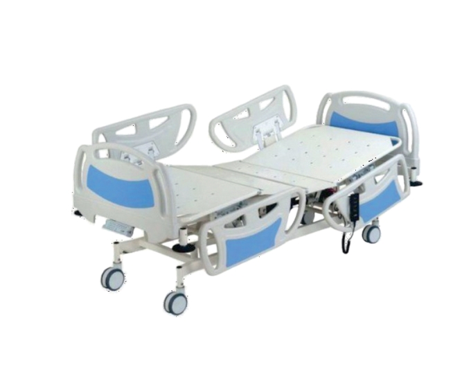
Electric ICU Bed with 5 Function System and ABS Side Rails
The Electric ICU Bed with 4 Motor System is a...
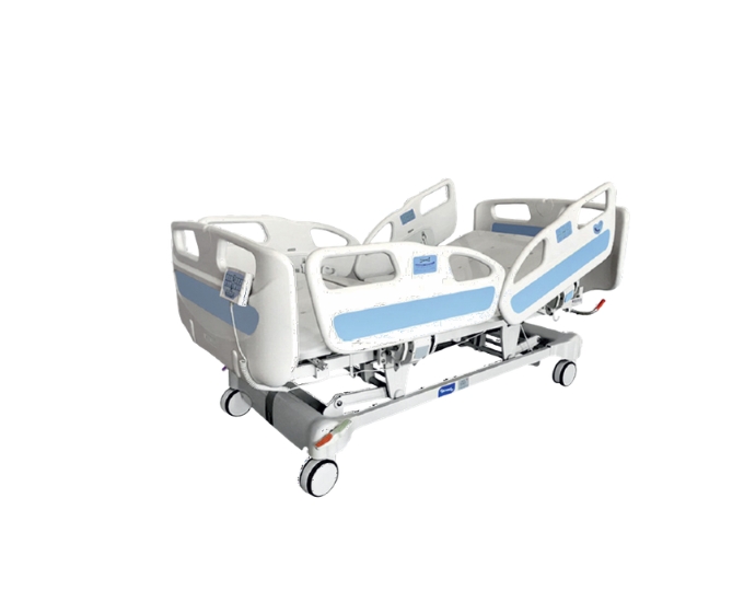
Electric ICU Bed with 4 Motor System and Trendelenburg Function
The Electric ICU Bed with 4 Motor System is a...
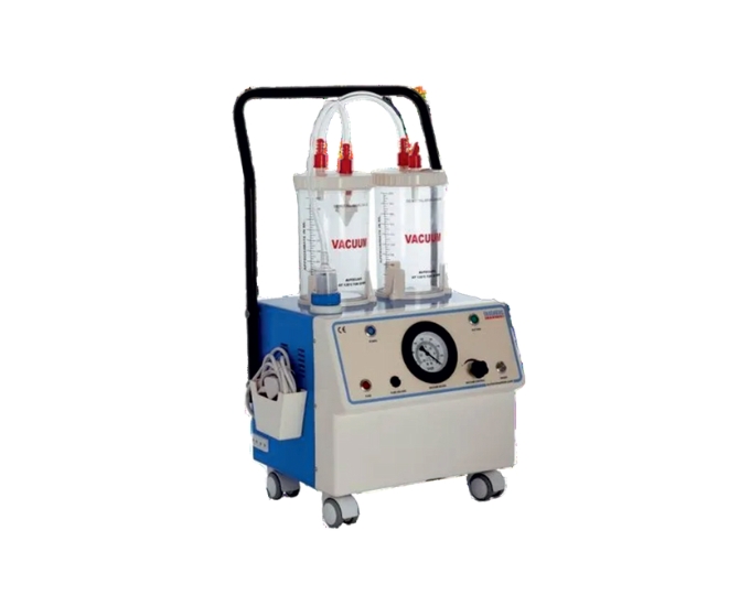
Twin Bottle Suction Machine with Vacuum Gauge and Oil Immersed Pump
The Twin Bottle Suction Machine is a reliable and efficient...
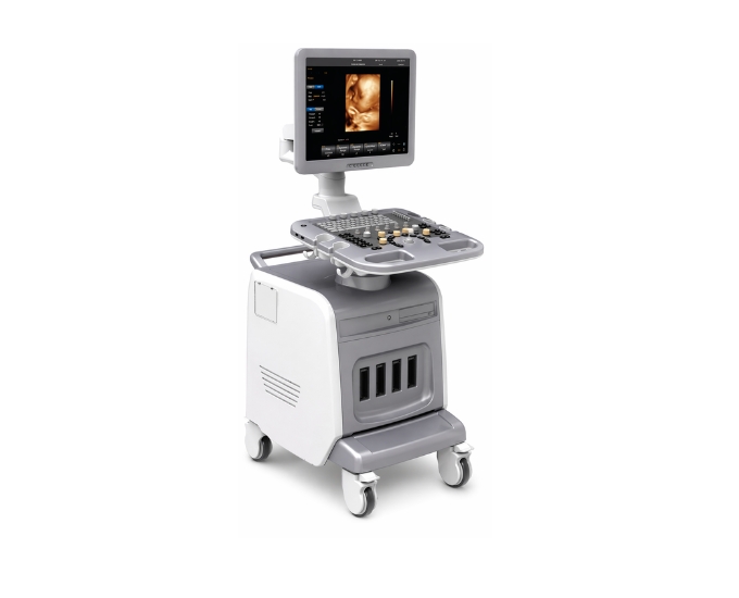
Advanced Color Doppler Ultrasound Diagnosis System for Precision Imaging
The Color Doppler Ultrasound Diagnosis System by Micro Technologies is...
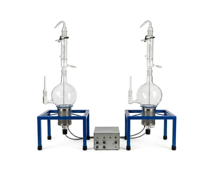
High-Performance NPL Type Distillation Apparatus
Micro Technologies offers a high-performance NPL type distillation apparatus designed...
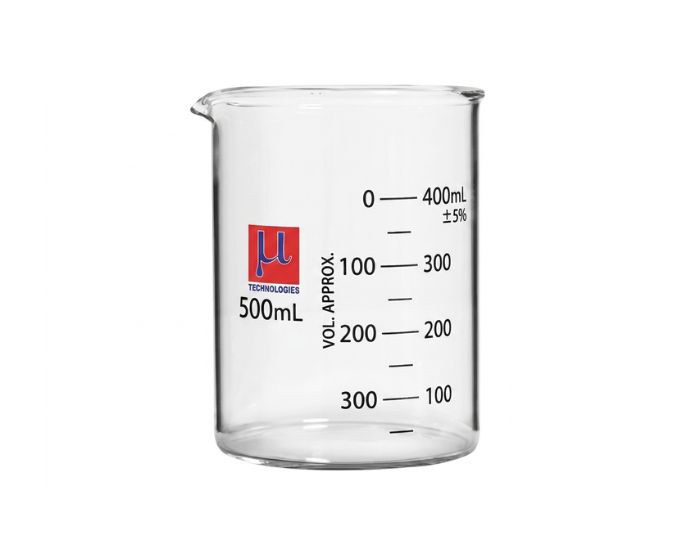
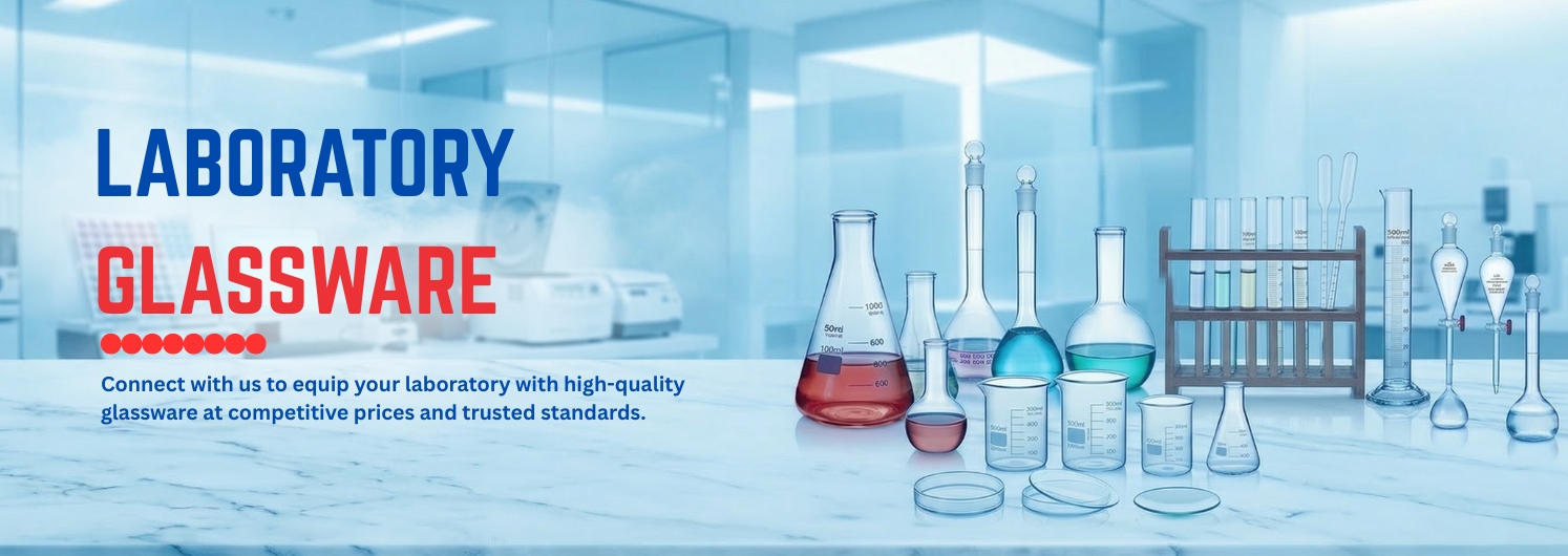
Low Form Beaker with Graduation for Laboratory Use
The Low Form Beaker with Graduation is a laboratory glassware...
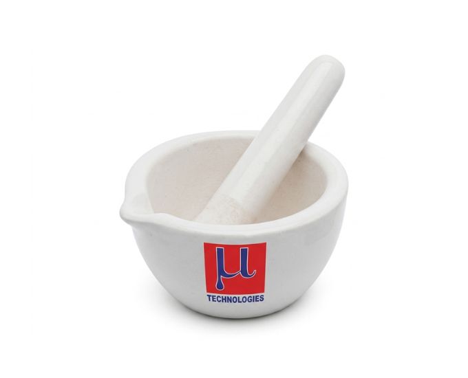
Pestle Mortar
Micro Technologies offers Pestle Mortar suitable for grinding, mixing, and...
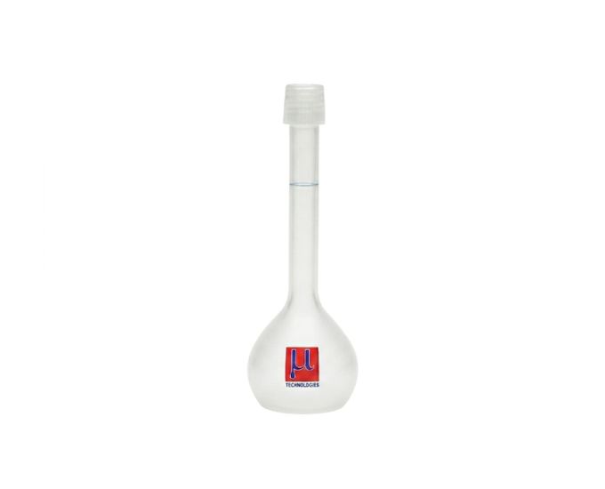
Plastic Volumetric Flask Class A
Plastic Volumetric Flask Class A is a precision laboratory measuring...
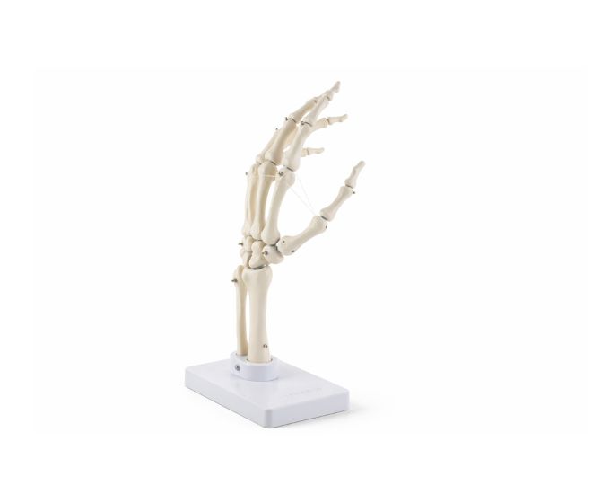
Hand joint without ligaments
Hand Joint without Ligaments is a life-size anatomical model designed...
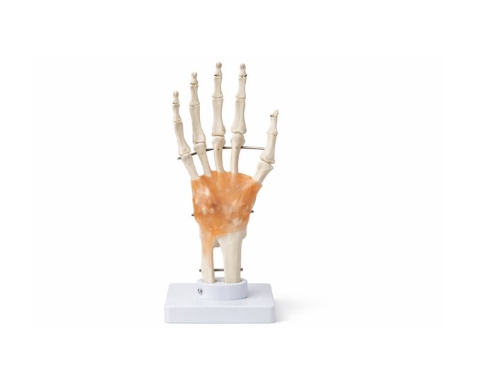
Hand joint With Ligament
Hand Joint with Ligament is a detailed anatomical model designed...
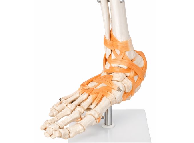
Foot joint life size With Ligaments
Foot Joint Life Size With Ligaments is a detailed anatomical...
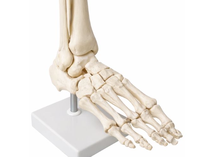
Foot joint Without Ligaments
The Foot Joint (Without Ligaments) anatomical model is a detailed...
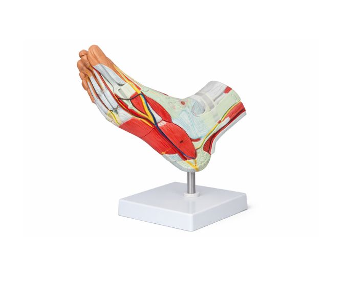
Muscle of Foot with Main Vessel and Nerve Model
The Human Foot Muscle Anatomy Model with Vessels and Nerves...
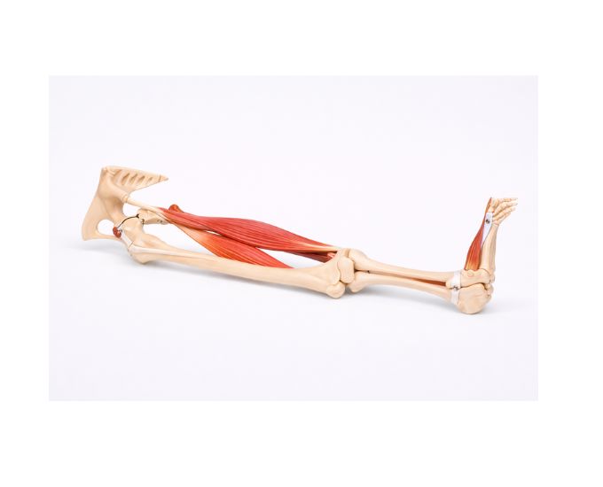
Functional model of knee joint life size
Functional Model of Knee Joint is a life-size anatomical model...
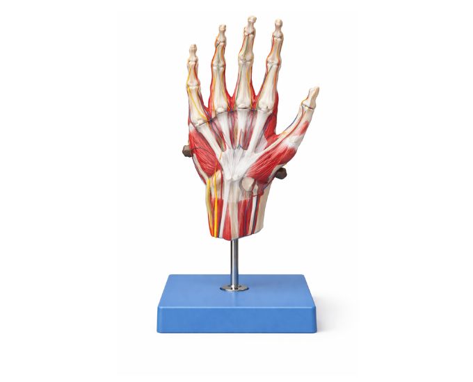
Muscle of hand with maine vessel and nerve
The Human Hand Muscle Anatomy Model with Vessels and Nerves...
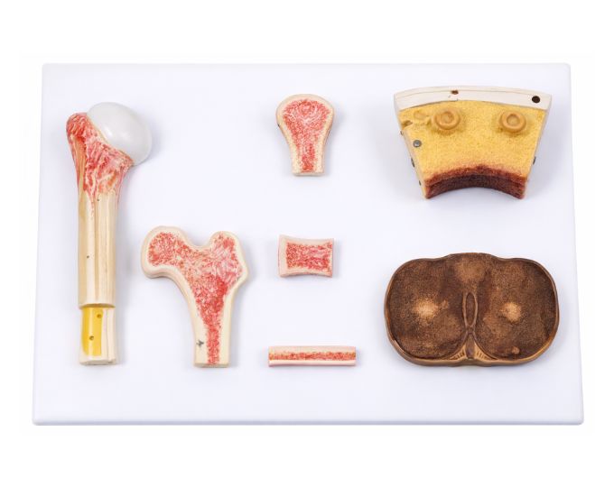
Bone tissue model life size
Bone Tissue Model Life Size is an anatomical teaching model...
Get in Touch With Us
Have questions about our products? Need customized solutions? Our team is here to assist you.
Laboratory Equipment
Premium quality lab equipment and glassware for all your research needs with certified standards.
Educational Solutions
Comprehensive educational products and kits designed for schools, colleges, and training institutions.
Custom Solutions
Tailored equipment packages and custom solutions built specifically for your unique requirements.
Expert Support
24/7 customer support with technical assistance from industry experts to help you succeed.
What Our Clients Say
Trusted by leading research institutions, educational centers, and laboratories across the globe.
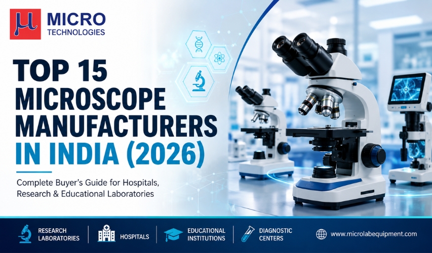
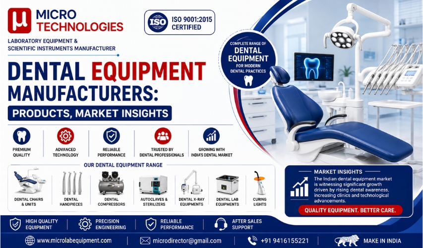
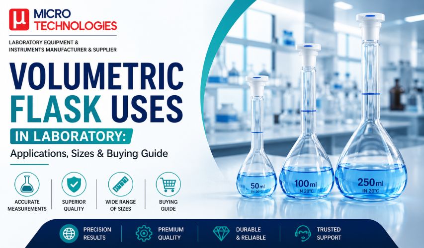
Latest Articles & Insights
Discover expert tips, guides, and industry updates from our laboratory equipment specialists.