
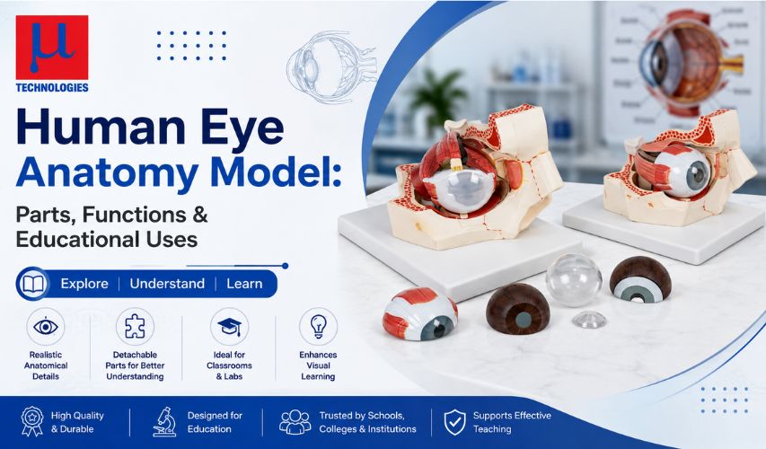
A human eye anatomy model is a physical (usually enlarged and dissectible) replica of the human eyeball used to teach the structure and function of vision. According to the National Eye Institute (NEI), light passes through the cornea, pupil, and lens before reaching the retina, where it is converted into electrical signals sent to the brain via the optic nerve. Educational eye models — typically enlarged 3x to 6x and made of durable PVC — are used in schools, biology and health science classrooms, optometry and ophthalmology training programs, and patient counseling to make this process easier to visualize.
The human eye is one of the most complex organs in the body, and understanding it from a textbook diagram alone can be difficult. That's where a human eye anatomy model becomes valuable—it turns a flat diagram into a hands-on, dissectible object that students and trainees can examine layer by layer. This guide covers the real anatomy the model represents, why enlarged and dissectible eye models are used in education, and how they support learning in schools, colleges, and clinical training settings—with every fact linked to its source.
What Is a Human Eye Anatomy Model?
A human eye anatomy model is a scaled-up, three-dimensional replica of the human eyeball, typically built with detachable or dissectible parts so learners can separate and examine individual structures. Commercial eye models sold for classroom and clinical use are commonly enlarged 3 to 6 times actual size and made of PVC plastic for durability, according to product listings from anatomy model suppliers such as Anatomy Warehouse and Samtech Instruments. One example, the Giant Eye Model manufactured by Micro Technologies (Ambala, India), is enlarged six times and mounted on a stand, with parts that separate to show the eye's outer covering, middle layer, retina, and internal lens structures.
The Real Anatomy Behind the Model
To understand what an eye anatomy model is showing, it helps to know the actual structure of the human eye, as documented by the National Eye Institute (NEI) and NCBI's StatPearls anatomy reference.
Size and Structure
The adult human eyeball measures about 2.5 centimeters (roughly 1 inch) in diameter, and only about one-sixth of it is visible from outside the body—the rest sits protected within the bony eye socket (orbit), according to StatPearls (NCBI Bookshelf, National Library of Medicine). Anatomically, the wall of the eyeball is made up of three layers, often called tunics:
- Fibrous tunic – the outer layer, made up of the transparent cornea at the front and the white sclera covering the rest of the eyeball.
- Vascular Tunic (Uvea): The nutrient-rich middle coat of the eye, made up of the choroid, ciliary body, and iris.
- Retina – the innermost, light-sensitive layer.
How Light Travels Through the Eye
According to the NEI, vision works in a specific sequence:
- Light first passes through the cornea, the clear, dome-shaped front layer that begins bending (refracting) light.
- Light then passes through the pupil, the opening whose size is controlled by the iris—the colored part of the eye.
- Just behind the iris, the lens performs secondary refraction to precisely sharpen the light path.
- Light reaches the retina at the back of the eye, where photoreceptor cells convert it into electrical signals.
- The optic nerve transmits these electrical impulses directly to the visual cortex, where the brain decodes them into recognizable sight.
The cornea does most of the eye's focusing work: according to the American Academy of Ophthalmology (AAO), a relaxed human eye has a total optical power of about 60 diopters, with the cornea alone contributing about 40 diopters—roughly two-thirds of the eye's total focusing power. The lens supplies the remaining adjustable portion, which is what allows the eye to shift focus between near and far objects.
Inside the Retina
The retina itself is layered, containing ganglion cells, bipolar cells, and photoreceptor cells, per StatPearls. There are two types of photoreceptors:
- Rods – roughly 100 million or more per retina, responsible for vision in dim light.
- Cones – about 6 million per retina, responsible for color vision.
At the center of the retina lies the macula lutea, and within it, the fovea centralis—the area containing only cones and responsible for the sharpest, most detailed vision. Where the optic nerve exits the eye, there are no photoreceptors at all; this point is known as the optic disc, or blind spot.
Fluids and Internal Chambers
Behind the iris, the lens divides the inside of the eyeball into two main spaces, as described by StatPearls:
- The anterior cavity (in front of the lens), filled with aqueous humor, a watery fluid produced by the ciliary body that nourishes the cornea and lens and is fully replaced roughly every 90 minutes.
- The vitreous cavity (behind the lens), filled with the vitreous humor, a clear, jelly-like substance that holds the retina in place against the choroid and does not get replaced quickly.
Muscles and Accessory Structures
Six extrinsic (extraocular) muscles move each eye: the superior rectus, inferior rectus, lateral rectus, medial rectus, superior oblique, and inferior oblique, as documented by StatPearls. Accessory structures — the eyelids, eyelashes, eyebrows, lacrimal (tear-producing) glands, and conjunctiva — protect the eye and keep its surface lubricated.
Why These Details Matter for a Physical Model
A well-designed human eye anatomy model reflects this real structure directly. For example, the Giant Eye Model from Micro Technologies is built to separate into parts that show the tunica externa (cornea and sclera, with attachments for the ocular muscles and optic nerve), the tunica media (iris, ciliary body, and choroid), the tunica interna (retina), and the refractive media (lens and vitreous body)—mirroring the three-tunic structure described in anatomical references. This kind of dissectible design lets a learner physically separate the same layers that textbooks describe only in cross-section diagrams.
Educational Uses of a Human Eye Anatomy Model
Based on how these models are marketed and used by suppliers such as 3B Scientific, Anatomy Warehouse, and Micro Technologies, human eye anatomy models serve several practical purposes in education and training:
1. Biology and Health Science Classrooms
Models help students visualize eye structures such as the cornea, lens, retina, optic nerve, and extraocular muscles—structures that are difficult to picture from a 2D diagram alone, according to 3B Scientific's product documentation.
2. Optometry and Ophthalmology Training
Eye models are used in optometry and ophthalmology training programs to give trainees a hands-on understanding of ocular anatomy, which supports learning how eye conditions are diagnosed and treated.
3. Patient Education
Some eye models are used during clinical consultations to help patients understand their own eye conditions and proposed treatments, since a physical model can improve comprehension compared to verbal explanation alone.
4. Demonstrating Common Eye Conditions
Certain models are designed specifically to help explain conditions such as cataracts and glaucoma, giving educators and clinicians a visual way to describe how these conditions affect the eye's structures.
5. Middle and High School STEM Activities
Some eye anatomy teaching tools are designed as labeling or construction activities for middle school students, combining hands-on assembly with vocabulary such as cornea, iris, lens, pupil, optic nerve, retina, sclera, and vitreous humor.
Choosing an Eye Anatomy Model for Teaching
When selecting a human eye anatomy model for a classroom, lab, or clinic, a few practical factors matter
- Magnification – Most educational eye models are enlarged 3x to 6x actual size, making small structures like the ciliary body and optic disc easier to see and point to.
- Number of dissectible parts – More separable parts generally allow more structures to be studied individually, though this also means more small pieces to manage in a classroom setting.
- Material and durability – PVC plastic construction is common for classroom models because it withstands repeated handling.
- Mounting – A model mounted on a stand, like the Micro Technologies Giant Eye Model, is easier to display, rotate, and pass around during a lesson.
Conclusion
A human eye anatomy model works because it mirrors real, well-documented anatomy—the three tunics of the eyeball, the path light takes from cornea to retina; the role of rods and cones; and the fluid-filled chambers that give the eye its shape, as described by the National Eye Institute, the American Academy of Ophthalmology, and NCBI's StatPearls reference. Whether used in a school biology class, an optometry training program, or a doctor's consultation room, a dissectible, enlarged eye model turns this anatomy into something students and patients can hold, take apart, and understand directly — which is why manufacturers such as Micro Technologies continue to supply these models to educational and medical institutions.
Frequently Asked Questions
What is a human eye anatomy model used for?
It is used to teach the structure and function of the human eye in biology and health science classrooms, optometry and ophthalmology training, and patient education, by letting learners see and separate the eye's internal layers.
What parts does a human eye model typically show?
Common educational eye models show the cornea, sclera, iris, lens, retina, optic nerve, ciliary body, choroid, and vitreous body, based on product documentation from anatomy model suppliers.
How much are eye anatomy models usually enlarged?
Most classroom and clinical eye models are enlarged between 3x and 6x actual size, since the real human eyeball is only about 2.5 centimeters in diameter, according to StatPearls (NCBI).
Why is the cornea considered so important in the eye's anatomy?
According to the American Academy of Ophthalmology, the cornea contributes about two-thirds of the eye's total focusing (refractive) power, making it central to how the eye forms a clear image.
Who manufactures human eye anatomy models?
Several companies manufacture these models, including Micro Technologies (Ambala, India), which produces a six-times-enlarged Giant Eye Model with detachable parts for educational use.