Muscle of Foot with Main Vessel and Nerve Model
Product Specification Sheet
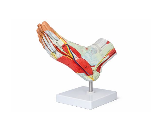
Muscle of Foot with Main Vessel and Nerve Model
Model Number: MT-030
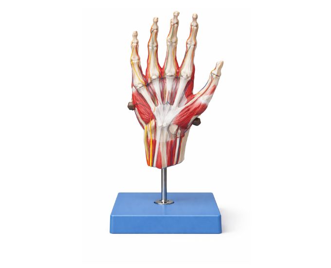
The Human Foot Muscle Anatomy Model with Vessels and Nerves is a detailed educational tool designed to demonstrate the internal structure of the human foot. It clearly displays muscles, blood vessels, nerves, and supporting ligaments, providing a comprehensive understanding of foot anatomy. Ideal for medical colleges, nursing institutes, physiotherapy training centers, and laboratories, this model supports effective teaching and learning. Manufactured by Micro Technologies, it features detachable parts for layer-by-layer study, helping students and professionals understand the functional anatomy of the lower limb with clarity and precision.
Product Specification
The Human Foot Muscle Anatomy Model with Vessels and Nerves is a highly detailed anatomical representation designed for in-depth study of the human foot. It accurately illustrates the muscular structure along with the main blood vessels, nerves, and ligamentous components that contribute to foot movement and stability. This model provides a clear understanding of how different structures interact to support balance and locomotion.
The model is divided into multiple detachable parts, allowing users to explore different layers of the foot step by step. This feature enhances learning by enabling a closer examination of individual components such as muscles, tendons, and neurovascular structures.
Crafted using high-quality materials, the model is durable and suitable for repeated use in classrooms and laboratories. Its realistic detailing and color coding make it easy to identify anatomical features, improving comprehension and retention.
Manufactured by Micro Technologies, this model is widely used in medical colleges, physiotherapy institutes, nursing schools, and research laboratories as an effective teaching and demonstration aid.
Features
- Detailed representation of foot muscles, vessels, and nerves
- Multiple detachable parts for layered study
- Clear visualization of ligament and structural components
- High-quality, durable construction
- Realistic design with accurate anatomical detailing
- Ideal for teaching and demonstration purposes
- Easy to assemble and disassemble
Applications
- Medical colleges
- Universities
- Laboratories
- Hospitals
- Research institutes
Product Specifications
| Specification | Details |
|---|---|
| Product Name | Human Foot Muscle Anatomy Model with Vessels and Nerves |
| Model No. | MT-030 |
| Type | Anatomical Model |
| Material | High-Quality PVC / Polymer |
| Size / Model | Standard Educational Model |
| Usage | Anatomy teaching and demonstration |
FAQs
Q1: What does this foot anatomy model display?
It shows the muscles, blood vessels, nerves, and ligaments of the human foot in a detailed and structured format.
Q2: Are the parts of this model detachable?
Yes, the model includes multiple removable parts that allow detailed study of different layers of the foot.
Q3: Who can use this anatomical model?
It is ideal for medical students, physiotherapy trainees, nursing students, and educators.
Q4: How does this model help in learning anatomy?
It provides a clear visual representation of foot structures, making it easier to understand their function and relationships.
Q5: Is the model durable for long-term use?
Yes, it is made from high-quality materials designed for repeated educational use.
Q6: Where is this model commonly used?
It is widely used in medical colleges, training institutes, laboratories, and research centers for teaching and demonstration.
Why Choose Our Products
- Manufactured by Micro Technologies with high anatomical accuracy
- Durable and long-lasting construction for repeated use
- Designed for clear visualization and easy understanding
- Ideal for both educational and professional training purposes