Osteoporosis bone model life size
Product Specification Sheet
Osteoporosis bone model life size
Model Number: MT-044
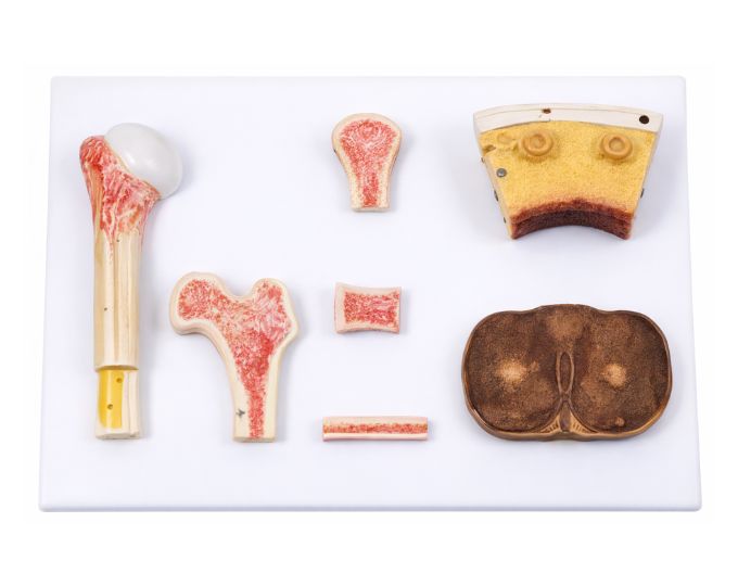
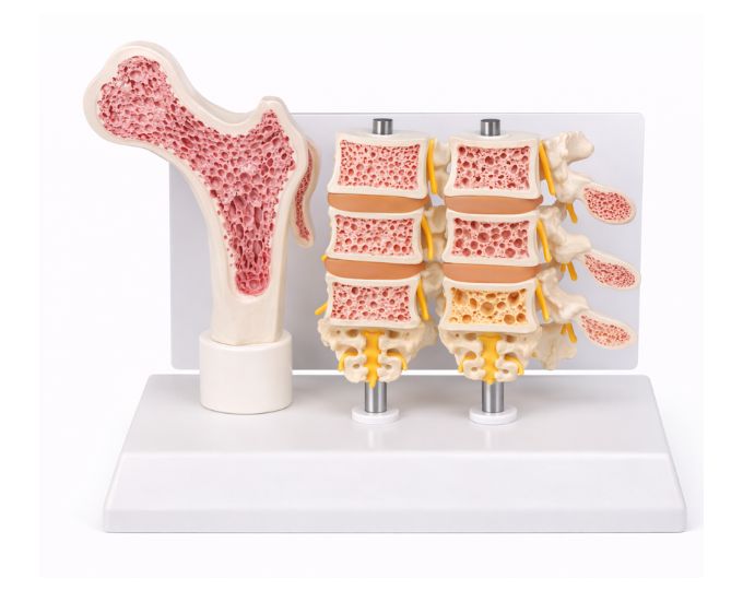
The Osteoporosis Bone Model is a life-size anatomical model designed to demonstrate the structural changes that occur in bones affected by osteoporosis. The model includes four bone sections, including three vertebrae, allowing students to clearly observe the effects of bone density loss and structural weakening. It helps learners understand how osteoporosis affects the internal structure of bones and increases the risk of fractures. By comparing healthy and osteoporotic bone structures, students can easily study the changes that occur in bone tissue during this condition. This anatomical model is widely used in medical colleges, nursing institutes, physiotherapy training centers and biology laboratories for teaching bone diseases and skeletal anatomy. The life-size design and clear anatomical representation make it an effective educational tool for classroom demonstrations and medical training.
Product Specification
The Life-Size Osteoporosis Bone Model with Vertebrae for Comparative Anatomy Study is an essential educational tool for understanding bone degeneration. Manufactured by Micro Technologies, this model includes four bone sections, including three vertebrae, to demonstrate the progressive effects of osteoporosis.
The Osteoporosis Bone Model Life Size allows students and healthcare professionals to visually compare healthy bone structure with osteoporotic changes. It clearly illustrates reduced bone density, structural weakening, and increased fracture risk, making complex concepts easier to understand.
Constructed with high-quality materials, the Osteoporosis Bone Model Life Size is durable and suitable for repeated classroom and laboratory use. It is widely used in medical colleges, nursing institutes, physiotherapy training centers, and biology labs. This model enhances teaching efficiency and supports practical learning. Contact Micro Technologies today to request a quote or explore bulk procurement options.
Features
- Life-size anatomical representation for realistic learning.
- Includes four bone sections including three vertebrae.
- Clear comparison of healthy vs osteoporotic bones.
- Detailed internal bone structure visualization.
- Durable construction for repeated educational use.
- Suitable for medical and anatomy training applications.
Benefits
- Enhanced Learning Experience: Simplifies understanding of osteoporosis.
- Improved Teaching Efficiency: Ideal for demonstrations and practical sessions.
- Accurate Visualization: Helps compare normal and diseased bone structures.
- Ease of Use: Lightweight and easy to display.
- Long-Term Value: Durable design ensures extended usability.
Product Specifications
| Specification | Details |
|---|---|
| Product Name | Osteoporosis Bone Model Life Size |
| Model No. | MT-044 |
| Type | Anatomical Teaching Model |
| Material | High-Quality PVC / Polymer |
| Size / Model | Life-Size Model |
| Usage | Bone disease education and anatomical study |
FAQs
What does the osteoporosis bone model demonstrate?
This model demonstrates the structural changes that occur in bones affected by osteoporosis, including reduced bone density and weakening of bone structure, helping users understand disease progression.
How does this model help in medical education?
It provides a clear visual comparison between healthy and osteoporotic bones, making it easier for students to understand bone degeneration and its impact on skeletal health.
Is the model suitable for classroom and laboratory use?
Yes, it is designed for use in medical colleges, nursing institutes, physiotherapy training centers, and biology laboratories for teaching and demonstrations.
Is the model durable for repeated handling?
Yes, it is made from high-quality materials that ensure durability and long-term usability in educational environments.
Does Micro Technologies provide bulk supply and customization?
Yes, Micro Technologies offers bulk procurement options, customization, and after-sales support for educational institutions and healthcare facilities.
Why Choose Our Products
- Quality: Designed to meet medical and educational standards.
- Durability: Built with robust materials for long service life.
- Accuracy & Performance: Ensures precise anatomical representation.
- Usability & Support: Easy-to-use design with reliable customer assistance.
Call to Action
Upgrade your anatomy training with this life-size osteoporosis bone model. Request a quote today or contact us for bulk orders and institutional pricing—trusted by educators and healthcare professionals.