Maxillary Teeth Caries Model 2 Parts for Dental Anatomy Study
Product Specification Sheet
Maxillary Teeth Caries Model 2 Parts for Dental Anatomy Study
Model Number: MT-110
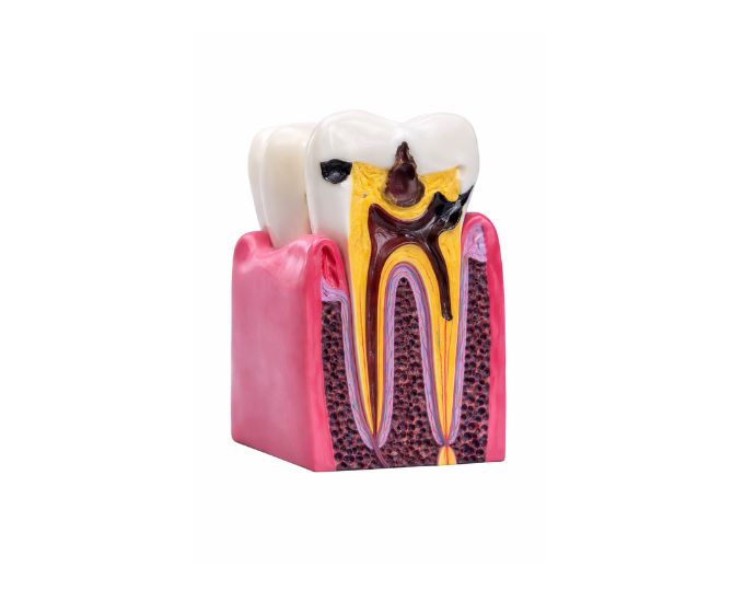
The Maxillary Teeth Caries Model is a dental teaching model designed to demonstrate the structure of a maxillary tooth and the development of dental caries. This model clearly illustrates the internal and external anatomy of a tooth affected by decay, helping students understand how caries progress through different layers of the tooth. The model consists of two detachable parts, allowing detailed observation of tooth structure. It is widely used in dental colleges, universities, medical institutes, and biology laboratories for dental anatomy and oral pathology education. This teaching model supports practical demonstrations and helps improve understanding of dental diseases.
Product Specification
The Maxillary Teeth Caries Model is a precision-designed educational tool developed to help students understand tooth anatomy and the development of dental caries. Created by Micro Technologies, this model clearly illustrates both the healthy structure of a maxillary tooth and the stages of decay affecting its layers.
The Maxillary Teeth Caries Model consists of 2 detachable parts, allowing learners to examine internal and external tooth structures in detail. This hands-on design helps students visualize how caries progress from enamel to deeper layers, improving understanding of oral pathology and preventive dentistry.
Widely used in dental colleges, universities, and biology laboratories, the Maxillary Teeth Caries Model is made from high-quality materials for long-term durability. It is an essential teaching aid for dental anatomy and oral disease education. Contact Micro Technologies today to request a quote or explore bulk purchasing options.
Features
- Demonstrates maxillary tooth structure and caries progression
- Clearly shows layers affected by dental decay
- 2 detachable parts for detailed anatomical study
- Accurate representation of tooth anatomy and pathology
- High-quality, durable PVC construction
- Ideal for dental education and laboratory use
- Suitable for classroom demonstrations and practical training
Benefits
- Enhances understanding of dental caries development
- Supports visual and hands-on learning in dentistry
- Cost-effective solution for educational institutions
- Improves clarity in teaching oral pathology concepts
- Easy to handle, assemble, and maintain
Product Specifications
| Specification | Details |
|---|---|
| Product Name | Maxillary Teeth Caries Model 2 Parts |
| Model No. | MT-110 |
| Type | Dental Anatomy & Pathology Teaching Model |
| Material | High-quality PVC |
| Size / Model | Standard Educational Size |
| Usage | Dental education, oral pathology training, laboratory demonstrations |
FAQs
What does the maxillary teeth caries model demonstrate?
This model demonstrates the structure of a maxillary tooth along with the progression of dental caries. It helps students understand how decay spreads through different layers of the tooth.
How does the 2-part design help in learning?
The 2 detachable parts allow students to examine both external and internal tooth structures separately. This makes it easier to understand tooth anatomy and the effects of decay in a practical way.
Is this model suitable for dental training programs?
Yes, the model is widely used in dental colleges, universities, and medical institutes. It is suitable for both basic and advanced dental anatomy and pathology education.
How durable is the model for regular use?
The model is made from high-quality PVC material, ensuring durability and resistance to wear. It is designed for long-term use in classrooms and laboratories while maintaining accuracy and detail.
Why Choose Our Products
- Quality: Manufactured to meet dental and medical education standards
- Durability: Strong construction ensures long service life
- Accuracy: Precise anatomical detailing for reliable learning
- Support: User-friendly design with dependable customer assistance