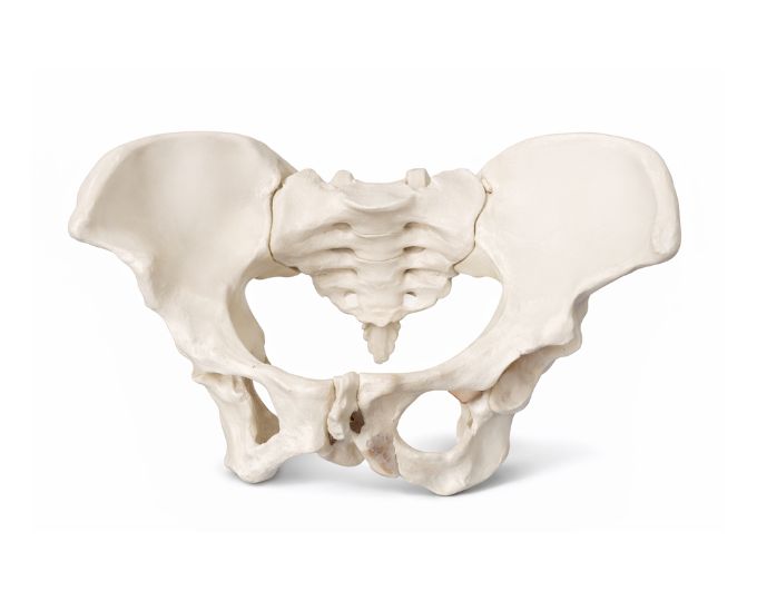
Lumbar Vertebra Sacrum Bone with Spinal Nerves Model
Product Specification Sheet
Lumbar Vertebra Sacrum Bone with Spinal Nerves Model
Model Number: MT-040
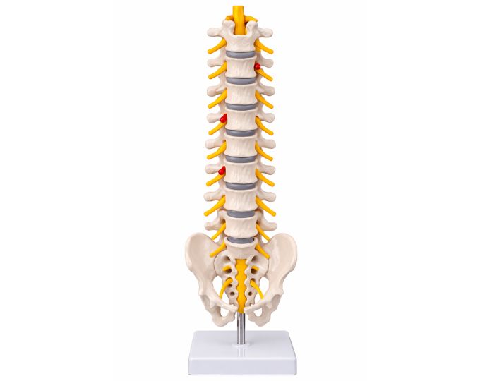
Lumbar Vertebra Sacrum Bone with Spinal Nerves is a detailed anatomical model designed to demonstrate the structure of the lower part of the human spine. The model clearly shows the lumbar vertebrae, sacrum and the spinal nerves emerging from the spinal column. It helps students understand the arrangement of the lower spinal bones and the pathway of spinal nerves that connect the spinal cord to different parts of the body. The model provides a clear visual reference for studying the anatomy of the lumbar region and the relationship between vertebrae and spinal nerves. This anatomical model is widely used in medical colleges, nursing institutes, physiotherapy training centers and biology laboratories for teaching spinal anatomy. The detailed structure and durable construction make it suitable for classroom demonstrations and practical learning.
Product Specification
The Detailed Lumbar Vertebra and Sacrum Model with Spinal Nerves for Anatomy Education is an essential educational tool for studying the lower section of the human spine. Manufactured by Micro Technologies, this model provides a clear and accurate representation of lumbar vertebrae, sacrum, and associated spinal nerves.
The Lumbar Vertebra Sacrum Bone with Spinal Nerves Model is designed to help students visualize the anatomical structure and relationships within the lower spinal region. It clearly displays how spinal nerves emerge from the vertebral column and connect to different parts of the body, aiding in comprehensive learning.
Built with durable materials, the Lumbar Vertebra Sacrum Bone with Spinal Nerves Model is suitable for repeated classroom demonstrations and hands-on learning. It is widely used in medical colleges, nursing institutes, physiotherapy training centers, and biology laboratories. This model enhances teaching efficiency and supports better conceptual understanding. Contact Micro Technologies today to request a quote or explore bulk procurement options.
Features
- Detailed anatomical representation of lumbar vertebrae and sacrum.
- Clearly visible spinal nerves for enhanced learning.
- Accurate structural design for educational use.
- Durable construction for repeated handling.
- Suitable for medical, nursing, and physiotherapy training.
- Compact and easy-to-display model for classrooms.
Benefits
- Improved Learning Experience: Helps students visualize spinal anatomy clearly.
- Enhanced Teaching Efficiency: Ideal for demonstrations and practical sessions.
- Accurate Knowledge Delivery: Supports detailed anatomical understanding.
- User-Friendly Design: Easy to handle and display.
- Long-Term Value: Durable build ensures extended usage.
Product Specifications
| Specification | Details |
|---|---|
| Product Name | Lumbar Vertebra Sacrum Bone with Spinal Nerves Model |
| Model No. | MT-040 |
| Type | Anatomical Teaching Model |
| Material | High-Quality PVC / Polymer |
| Size / Model | Standard Educational Model |
| Usage | Anatomy education and demonstration |
FAQs
What is this anatomical model used for?
This model is used to demonstrate the structure of the lumbar spine, sacrum, and spinal nerves. It helps students and educators understand the arrangement and function of the lower spinal region.
Is the model accurate for medical education?
Yes, the model is designed with high anatomical accuracy to support medical, nursing, and physiotherapy education, making it suitable for professional training.
Can this model be used in classrooms and laboratories?
Absolutely, it is ideal for classroom demonstrations, laboratory studies, and practical training sessions across educational institutions.
Is the model durable for frequent use?
Yes, it is made from high-quality materials that ensure durability and long-term use even with regular handling.
Does Micro Technologies offer bulk supply for institutions?
Yes, Micro Technologies provides bulk supply options, customization, and after-sales support for educational institutions and laboratories.
Why Choose Our Products
- Quality: Designed to meet educational and medical standards.
- Durability: Made from robust materials for long-term use.
- Accuracy & Performance: Provides precise anatomical representation.
- Usability & Support: Easy-to-use design with reliable customer assistance.
Call to Action
Enhance your anatomy teaching tools with this detailed spine model. Request a quote today or contact us for bulk orders and institutional pricing—trusted by educators and professionals.