Human Eye Model with Pathological Cornea
Product Specification Sheet
Human Eye Model with Pathological Cornea
Model Number: MT-088
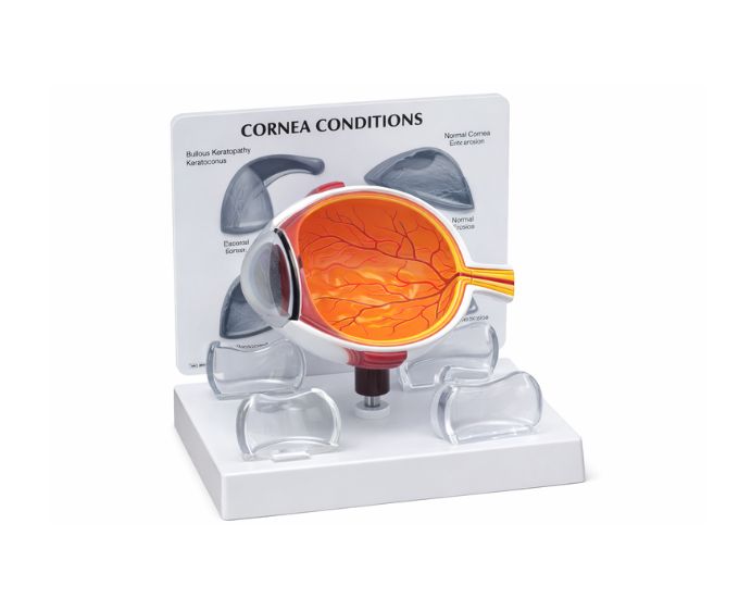
The Human Eye Model with Pathological Cornea is an educational anatomical model designed to demonstrate the structure of the human eye along with common corneal pathologies. This model helps students understand how diseases and abnormalities can affect the cornea and influence vision. The model clearly illustrates the anatomical structure of the eye and highlights pathological changes in the cornea, making it easier for learners to study different eye conditions. An instructional card is included with the model to explain the displayed corneal pathologies and anatomical details. This visual and interactive design helps students better understand eye diseases and their impact on the visual system. The model is widely used in medical colleges, nursing institutes, ophthalmology training centers and biology laboratories for teaching eye anatomy and eye pathology.
Product Specification
The Human Eye Anatomy Model with Pathological Cornea is an advanced teaching aid developed to provide a comprehensive understanding of eye anatomy and corneal diseases. Designed by Micro Technologies, this model clearly illustrates the structure of the human eye along with visible pathological changes in the cornea.
The Human Eye Anatomy Model with Pathological Cornea enables students to study how various corneal conditions impact vision and eye function. It highlights key anatomical features while also demonstrating abnormalities, helping learners bridge the gap between theory and clinical understanding. The included instructional card further enhances learning by explaining the displayed pathologies in detail.
Widely used in medical colleges, nursing institutes, ophthalmology training centers, and laboratories, the Human Eye Anatomy Model with Pathological Cornea is made from durable materials for long-term use. It is an essential tool for teaching ophthalmic anatomy and pathology. Contact Micro Technologies today to request a quote or discuss bulk purchasing options.
Features
- Demonstrates both normal eye anatomy and corneal pathologies
- Clearly displays corneal abnormalities affecting vision
- Includes instructional card for detailed explanation
- Accurate anatomical detailing for effective learning
- High-quality, durable PVC construction
- Ideal for ophthalmology and medical training
- Suitable for classroom and laboratory demonstrations
Benefits
- Enhances understanding of eye diseases and corneal conditions
- Supports visual and clinical learning experiences
- Cost-effective solution for medical and educational institutions
- Improves clarity in teaching ophthalmic pathology
- Easy to use, handle, and maintain
Product Specifications
| Specification | Details |
|---|---|
| Product Name | Human Eye Model with Pathological Cornea |
| Model No. | MT-088 |
| Type | Anatomical & Pathology Teaching Model |
| Material | High-quality PVC |
| Size / Model | Standard Educational Size |
| Usage | Ophthalmology education, pathology training, laboratory demonstrations |
FAQs
What types of corneal pathologies are shown in this model?
This model illustrates common corneal abnormalities that affect vision, helping students understand how diseases alter the structure and function of the eye. The included instructional card provides detailed explanations of each condition.
Is this model suitable for ophthalmology training?
Yes, the model is widely used in ophthalmology training centers, medical colleges, and nursing institutes. It provides both anatomical and pathological insights, making it suitable for comprehensive learning.
How does this model improve understanding of eye diseases?
By visually demonstrating corneal pathologies alongside normal anatomy, the model helps students compare healthy and diseased conditions. This enhances understanding of how eye disorders develop and impact vision.
Is the model durable for long-term educational use?
The model is made from high-quality PVC material, ensuring durability and resistance to wear. It is designed for repeated use in classrooms and laboratories while maintaining its accuracy and structural integrity.
Why Choose Our Products
- Quality: Manufactured to meet medical and educational standards
- Durability: Strong construction ensures long service life
- Accuracy: Precise anatomical detailing for reliable learning
- Support: User-friendly design with dependable customer assistance