Eyeball Imaging Demonstrator Model
Product Specification Sheet
Eyeball Imaging Demonstrator Model
Model Number: MT-095
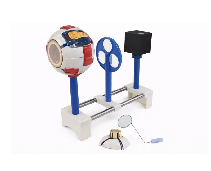
The Eyeball Imaging Demonstrator MT-095 is an educational model designed to demonstrate how images are formed within the human eye. It helps students understand the basic principles of vision, including how light passes through the eye and focuses on the retina. This model is widely used in medical colleges, universities, and biology laboratories for teaching eye anatomy and optical functions of the eye. By visually representing the process of image formation, the model simplifies complex concepts related to human vision. It is an effective teaching aid for instructors explaining the relationship between eye structure and visual perception in educational and laboratory environments.
Product Specification
The Eyeball Imaging Demonstrator Model – Vision & Optics Teaching Aid from Micro Technologies is developed to simplify the understanding of visual processes and eye optics. This model effectively demonstrates how light enters the eye, passes through different structures, and forms an image on the retina, helping students grasp fundamental concepts of human vision.
This Eyeball Imaging Demonstrator Model is widely used in medical colleges, universities, and biology laboratories where both anatomical and optical functions of the eye are taught. It bridges the gap between theory and practical learning by providing a clear, visual representation of image formation. The model is particularly useful for explaining the relationship between eye structure and visual perception in an engaging manner.
With durable construction and precise design, Micro Technologies ensures that this Eyeball Imaging Demonstrator Model delivers consistent performance and long-term reliability for professional educational environments.
Features
- Demonstrates image formation within the human eye
- Clearly explains light passage and focusing mechanism
- Accurate representation of eye optics and vision principles
- Durable construction for long-term educational use
- Ideal for classroom demonstrations and lab teaching
- Designed for medical and biology education environments
Benefits
- Simplifies complex vision and optics concepts for students
- Enhances teaching effectiveness with visual demonstrations
- Cost-effective tool for repeated academic use
- Supports safe and practical learning environments
- Provides long-term value with minimal maintenance
Product Specifications
| Specification | Details |
|---|---|
| Product Name | Eyeball Imaging Demonstrator Model |
| Model No. | MT-095 |
| Type | Anatomical & Optical Teaching Model |
| Material | High-Quality Durable PVC |
| Size / Model | Standard Educational Model |
| Usage | Medical Education, Laboratories, Training Institutes |
FAQs
What does the eyeball imaging demonstrator model explain?
This model explains how images are formed inside the human eye by demonstrating the path of light and how it focuses on the retina. It helps students understand key concepts of vision, including refraction and image formation, in a clear and visual way.
Is this model suitable for teaching both anatomy and optics?
Yes, the model is specifically designed to bridge anatomy and optical principles. It provides a combined understanding of eye structure and visual function, making it ideal for interdisciplinary teaching in medical and biology education.
Can this model be used in regular classroom demonstrations?
The model is built for frequent use in classrooms and laboratories. Its durable construction ensures that it can withstand repeated handling while maintaining its effectiveness as a teaching tool.
Do you offer bulk supply for educational institutions?
Micro Technologies provides bulk supply options for colleges, laboratories, and training institutes. Institutions can request customized quantities and receive competitive pricing by contacting the team directly.
Why Choose Our Products
- Quality Assurance: Designed to meet educational and scientific standards
- Durability: Built for long-lasting use in academic environments
- Accuracy: Precisely demonstrates vision and optical principles
- Ease of Use & Support: User-friendly design with reliable customer support
Request a quote today or contact us for bulk orders to upgrade your institution with advanced vision and anatomy teaching models.