Eye in Orbit Anatomy Model 8 Parts
Product Specification Sheet
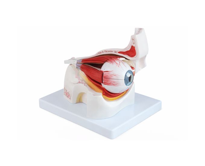
Eye in Orbit Anatomy Model 8 Parts
Model Number: MT-084
The Eye in Orbit Anatomy Model is a detailed educational model designed to demonstrate the structure of the human eye within the orbital cavity. This anatomical model clearly shows how the eye is positioned inside the eye socket and how it is supported by surrounding anatomical structures. The model helps students study important parts of the eye such as the eyeball, optic nerve and surrounding tissues that support vision and eye movement. With 8 detachable parts, learners can remove and examine individual components to better understand the structure of the eye and its relationship with the orbital cavity. The model provides a clear visual representation of eye anatomy, making it easier for students to study the human visual system. It is widely used in medical colleges, nursing institutes, biology laboratories and educational institutions for teaching ophthalmic anatomy.
Product Specification
The Eye in Orbit Anatomy Model is a high-quality educational tool designed to provide a detailed understanding of the human eye within its natural anatomical position. Developed by Micro Technologies, this model clearly illustrates the eyeball, optic nerve, and surrounding orbital structures that support vision and eye movement.
With 8 detachable parts, the Eye in Orbit Anatomy Model allows students to remove and examine individual components, helping them understand the structural relationships between the eye and the orbital cavity. This hands-on approach improves comprehension of ophthalmic anatomy and enhances learning outcomes.
Widely used in medical colleges, nursing institutes, and biology laboratories, the Eye in Orbit Anatomy Model is constructed from durable materials to ensure long-term use. It is an essential teaching aid for studying the human visual system and eye anatomy. Contact Micro Technologies today to request a quote or explore bulk purchasing options.
Features
- 8 detachable parts for detailed anatomical study
- Clearly shows eye position within the orbital cavity
- Displays eyeball, optic nerve, and supporting structures
- Accurate anatomical representation for effective learning
- High-quality, durable PVC construction
- Ideal for ophthalmic and anatomy education
- Suitable for classroom and laboratory demonstrations
Benefits
- Enhances understanding of eye anatomy and orbital structure
- Supports interactive and hands-on learning experiences
- Cost-effective solution for educational institutions
- Improves clarity in teaching ophthalmic concepts
- Easy to assemble, disassemble, and maintain
Product Specifications
| Specification | Details |
|---|---|
| Product Name | Eye in Orbit Anatomy Model 8 Parts |
| Model No. | MT-084 |
| Type | Anatomical Teaching Model |
| Material | High-quality PVC |
| Size / Model | Standard Educational Size |
| Usage | Ophthalmic education, laboratory training, classroom demonstrations |
FAQs
What does the “eye in orbit” feature demonstrate?
This model demonstrates how the eye is positioned within the orbital cavity and how it is supported by surrounding tissues. It helps students understand the relationship between the eyeball, optic nerve, and the structures that enable eye movement and protection.
Is this model suitable for ophthalmology training?
Yes, the model is widely used in medical colleges and nursing institutes for teaching ophthalmic anatomy. It provides accurate representation, making it suitable for both basic and advanced levels of study.
Can individual parts be removed for detailed study?
Yes, the model includes 8 detachable parts that can be removed and examined individually. This feature enhances hands-on learning and allows students to study each component in detail.
How durable is the model for regular use?
The model is made from high-quality PVC material, ensuring durability and resistance to wear. It is designed for long-term use in classrooms and laboratories while maintaining its structural accuracy and detail.
Why Choose Our Products
- Quality: Manufactured to meet medical and educational standards
- Durability: Strong construction ensures long service life
- Accuracy: Precise anatomical detailing for reliable learning
- Support: User-friendly design with dependable customer assistance