Eye Ball and Orbit with Vessels and Nerves 9 Parts
Product Specification Sheet
Eye Ball and Orbit with Vessels and Nerves 9 Parts
Model Number: MT-086
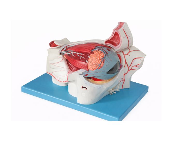
The Eye Ball and Orbit with Vessels and Nerves Model (MT-086) is a detailed anatomical teaching model designed to demonstrate the structure of the human eye and its surrounding orbital components. The model clearly illustrates the eyeball, blood vessels, and nerves, allowing students to study the complex anatomy of the visual system. Designed in 9 detachable parts with a size of 280 × 320 × 415 mm, it provides a comprehensive view of the orbital structure and eye anatomy. This model is widely used in medical colleges, biology laboratories, ophthalmology training, and educational institutions to support the study of human eye anatomy and visual system structure.
Product Specification
The Eye Ball and Orbit Anatomy Model with Vessels and Nerves is a precision-engineered educational tool developed to provide a detailed understanding of the human visual system. Designed by Micro Technologies, this model clearly illustrates the eyeball along with surrounding orbital structures, including blood vessels and nerves.
With 9 detachable parts, the Eye Ball and Orbit Anatomy Model with Vessels and Nerves allows students to remove and examine individual components, helping them understand the relationship between the eye, vascular system, and neural pathways. Its large size (280 × 320 × 415 mm) ensures excellent visibility, making it ideal for classroom demonstrations and laboratory teaching.
Widely used in medical colleges, biology laboratories, and ophthalmology training institutes, the Eye Ball and Orbit Anatomy Model with Vessels and Nerves is built from durable materials for long-term use. Contact Micro Technologies today to request a quote or discuss bulk purchasing options.
Features
- 9 detachable parts for detailed anatomical study
- Clearly displays eyeball, blood vessels, and nerves
- Large size (280 × 320 × 415 mm) for enhanced visibility
- Accurate anatomical detailing for effective learning
- High-quality, durable PVC construction
- Ideal for ophthalmology and anatomy education
- Suitable for classroom and laboratory demonstrations
Benefits
- Enhances understanding of eye anatomy and neural connections
- Supports interactive and hands-on learning experiences
- Cost-effective solution for educational institutions
- Improves clarity in teaching complex visual system structures
- Easy to assemble, disassemble, and maintain
Product Specifications
| Specification | Details |
|---|---|
| Product Name | Eye Ball and Orbit with Vessels and Nerves 9 Parts |
| Model No. | MT-086 |
| Type | Anatomical Teaching Model |
| Material | High-quality PVC |
| Size / Model | 280 × 320 × 415 mm |
| Usage | Ophthalmology education, laboratory training, anatomy demonstrations |
FAQs
What anatomical features are included in this model?
This model clearly displays the eyeball along with surrounding orbital structures such as blood vessels and nerves. It helps students understand how these components interact within the visual system.
Is this model suitable for advanced ophthalmology training?
Yes, the model is widely used in medical colleges and ophthalmology training institutes. Its detailed structure and large size make it suitable for both basic and advanced levels of study.
Can the model be disassembled for detailed examination?
Yes, the model consists of 9 detachable parts that can be removed individually. This feature supports hands-on learning and allows detailed examination of each anatomical component.
How durable is the model for long-term use?
The model is made from high-quality PVC material, ensuring durability and resistance to wear. It is designed for frequent use in classrooms and laboratories while maintaining its structural accuracy and detail.
Why Choose Our Products
- Quality: Manufactured to meet medical and educational standards
- Durability: Strong construction ensures long service life
- Accuracy: Precise anatomical detailing for reliable learning
- Support: User-friendly design with dependable customer assistance