Dissection Model of Eyepit and Periocular Anatomy
Product Specification Sheet
Dissection Model of Eyepit and Periocular Anatomy
Model Number: MT-094
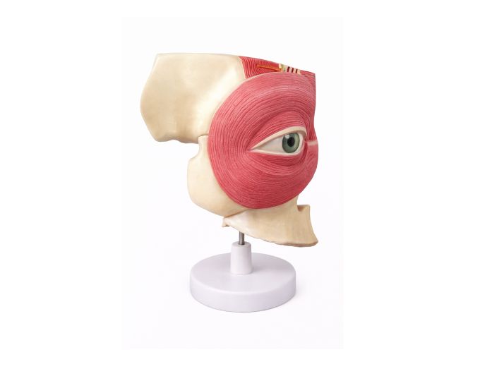
The Dissection Model of Eyepit and Periocular Anatomy MT-094 is a highly detailed anatomical teaching model designed to demonstrate the structure of the eye socket and surrounding tissues. Enlarged six times the natural size, the model clearly illustrates the periocular region including muscles, nerves, and related structures around the eye. It separates into six parts, allowing students and instructors to study the internal arrangement of the orbital cavity and its anatomical relationships. This model is widely used in medical colleges, universities, biology laboratories, and ophthalmology training institutes to support anatomy education and practical demonstrations.
Product Specification
The Advanced Eyepit & Periocular Anatomy Dissection Model – 6-Part Enlarged from Micro Technologies is engineered for accurate and comprehensive anatomical education. Enlarged six times its natural size, this model clearly demonstrates the intricate structures of the orbital cavity, including muscles, nerves, and surrounding periocular tissues.
This Eyepit & Periocular Anatomy Model features six detachable parts, enabling detailed examination of internal arrangements and spatial relationships within the eye socket. It is widely used in medical colleges, biology laboratories, and ophthalmology training centers where precision and clarity are essential. The model supports effective teaching of complex anatomical concepts and enhances hands-on learning experiences.
Built with durable materials, Micro Technologies ensures that this Eyepit & Periocular Anatomy Model delivers long-term reliability and consistent performance, making it a valuable addition to professional educational environments.
Features
- Enlarged 6x size for superior anatomical clarity
- Six detachable parts for detailed dissection study
- Clearly displays orbital cavity, muscles, and nerves
- Accurate representation of periocular structures
- Durable construction for long-term institutional use
- Ideal for ophthalmology and anatomy education
- Easy assembly and handling for repeated demonstrations
Benefits
- Enhances understanding of complex orbital anatomy
- Improves teaching efficiency with clear visual demonstration
- Cost-effective solution for long-term academic use
- Enables safe, hands-on learning without real dissection
- Provides reliable performance with minimal maintenance
Product Specifications
| Specification | Details |
|---|---|
| Product Name | Eyepit & Periocular Anatomy Model (6 Part) |
| Model No. | MT-094 |
| Type | Anatomical Dissection Model |
| Material | High-Quality Durable PVC |
| Size / Model | 6 Times Enlarged, 6 Detachable Parts |
| Usage | Medical Education, Laboratories, Training Institutes |
FAQs
What does the eyepit and periocular anatomy model demonstrate?
This model demonstrates the detailed structure of the orbital cavity along with surrounding periocular tissues such as muscles and nerves. Its enlarged design and detachable parts allow users to clearly study anatomical relationships, making it highly effective for teaching and practical demonstrations in medical education.
Is this model suitable for ophthalmology training?
Yes, the model is specifically designed for ophthalmology and advanced anatomy training. It provides accurate representation of the eye socket and related structures, making it ideal for use in medical colleges, training institutes, and specialized courses.
Can the model withstand regular use in institutions?
The model is constructed using durable, high-quality materials that ensure long-term performance even with frequent handling. It is designed to maintain structural integrity and accuracy, making it a reliable choice for classrooms and laboratories.
Is bulk ordering available for institutions?
Micro Technologies offers flexible bulk supply options for educational and healthcare institutions. Buyers can request customized quantities and receive competitive pricing by contacting the team directly for a quotation.
Why Choose Our Products
- Quality Assurance: Designed to meet medical and educational standards
- Durability: Strong construction ensures long-lasting usability
- Accuracy: Precisely crafted for realistic anatomical representation
- Ease of Use & Support: User-friendly design with reliable assistance
Request a quote today or contact us for bulk orders to equip your institution with advanced anatomical teaching models.