3D Human Vision System Wall-Mounted Anatomy Chart Model for Teaching
Product Specification Sheet
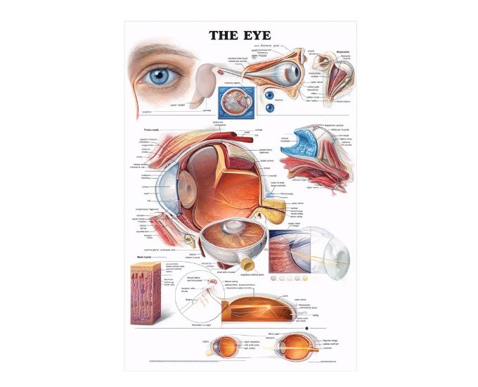
3D Human Vision System Wall-Mounted Anatomy Chart Model for Teaching
Model Number: MT-238
The 3D human vision system wall-mounted model is an advanced educational tool designed to demonstrate the anatomy of the human eye and visual system. It provides a detailed and visually engaging representation of eye structures and vision mechanisms. Ideal for medical colleges, universities, and training institutes, this model enhances classroom teaching and laboratory learning. Its wall-mounted design makes it perfect for display and demonstration, allowing easy visualization for groups. This model is highly useful for improving understanding of complex visual processes and anatomical relationships within the human eye.
Product Specification
The 3D human vision system wall-mounted anatomy model is a comprehensive educational aid designed to illustrate the structure and function of the human visual system. It features detailed representations of the eye, including internal components such as the retina, lens, optic nerve, and surrounding structures, helping users understand how vision occurs.
This model is crafted with precision using high-quality materials to ensure durability and long-term use in academic and clinical environments. The three-dimensional design enhances depth perception and clarity, making complex anatomical concepts easier to understand. Its wall-mounted format allows for convenient display in classrooms and laboratories, making it ideal for group teaching sessions.
Widely used in medical education, this model supports the study of ophthalmology and general human anatomy. It is an essential teaching aid for demonstrating visual pathways and eye structure, helping students and professionals gain a deeper understanding of the human vision system.
Features
-
Detailed 3D representation of the human vision system
-
Clearly illustrates eye anatomy and visual pathways
-
Wall-mounted design for easy display and teaching
-
Durable and high-quality construction
-
Large size for clear group visibility
-
Ideal for classroom and laboratory demonstrations
-
Enhances understanding of complex visual processes
Applications
-
Medical colleges
-
Universities
-
Laboratories
-
Hospitals
-
Research institutes
Product Specifications
| Specification | Details |
|---|---|
| Product Name | Vision System, Wall-Mounted, 3D |
| Model No. | MT-238 |
| Type | Anatomical Wall-Mounted Teaching Model |
| Material | High-quality PVC / Polymer |
| Size / Model | 770 × 550 × 10 mm |
| Usage | Medical education and anatomical demonstration |
FAQs
Q1. What does the 3D vision system model show?
It demonstrates the structure of the human eye and the functioning of the visual system in detail.
Q2. Is the model suitable for classroom teaching?
Yes, its wall-mounted design and large size make it ideal for group teaching and demonstrations.
Q3. Where can this model be used?
It is widely used in medical colleges, laboratories, hospitals, and training institutes.
Q4. What material is used in the model?
The model is made from durable, high-quality polymer materials for long-term use.
Why Choose Our Products
-
High anatomical accuracy for effective learning
-
Durable and long-lasting construction
-
Designed for clear visual demonstration
-
Suitable for educational and clinical environments