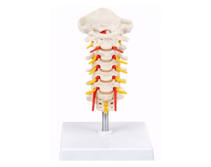
Occipital cervical vertebrae with vertebral artery model
Short Description (≈750–800 characters) The Model of Occipital, Cervical Vertebrae and Vertebral Artery is a life-size anatomical teaching model designed to demonstrate the structure of the upper part of the human spine and its connection with the skull. The model clearly shows the occipital bone, cervical vertebrae, vertebral artery, spinal nerves and brainstem. It helps students understand how the cervical vertebrae support the head and allow movement of the neck while protecting the spinal cord. The model also illustrates the pathway of the vertebral artery and spinal nerves in the cervical region, providing a clear understanding of blood supply and nerve distribution in this area. This anatomical model is widely used in medical colleges, nursing institutes, physiotherapy training centers and biology laboratories for teaching human anatomy and neuroanatomy.
Product Description
Occipital Cervical Vertebrae with Vertebral Artery Model
The Occipital Cervical Vertebrae with Vertebral Artery model is designed to provide a clear and detailed representation of the upper spinal region and its connection with the skull. This life-size anatomical model demonstrates the occipital bone, cervical vertebrae, vertebral artery, spinal nerves and brainstem.
The model helps learners understand how the skull connects to the cervical spine and how important structures such as blood vessels and nerves pass through this region. It provides an effective visual aid for studying the anatomy of the neck and the upper spinal column.
This model is widely used in medical colleges, physiotherapy institutes, nursing schools and anatomy laboratories as an educational tool for teaching spinal and neurological anatomy.
Anatomical Structure of the Model
Occipital Bone Structure
The model clearly shows the occipital bone located at the back of the skull, which connects the skull to the cervical vertebrae.
Cervical Vertebrae Representation
It displays the cervical vertebrae that form the upper part of the spinal column and support the head while allowing neck movement.
Vertebral Artery Pathway
The model demonstrates the vertebral artery passing through the cervical vertebrae, helping students understand the blood supply to the brain.
Spinal Nerves and Brainstem
The spinal nerves and brainstem are clearly represented, allowing learners to study their relationship with the cervical spine and skull.
Life Size Educational Model
The life-size design provides a realistic anatomical reference, making it easier for students to study the natural structure and arrangement of these components.
Product Specifications
| Specification | Details |
|---|---|
| Model Number | MT-042 |
| Product Name | Occipital Cervical Vertebrae with Vertebral Artery Model |
| Size | Life Size |
| Structure | Occipital Bone, Cervical Vertebrae, Vertebral Artery, Spinal Nerves and Brainstem |
| Material | High Quality PVC |
| Category | Anatomy Educational Model |
| Usage | Medical, Nursing and Biology Education |
| Application | Classroom Teaching, Laboratory Demonstration |
Frequently Asked Questions (FAQ)
What does this anatomical model show?
The model demonstrates the occipital bone, cervical vertebrae, vertebral artery, spinal nerves and brainstem.
Is the model life size?
Yes, the model is designed in life-size proportion for realistic anatomical study.
Who can use this anatomical model?
Medical students, nursing students, physiotherapy trainees and anatomy instructors commonly use this model.
Where is this model used?
It is widely used in medical colleges, physiotherapy institutes, nursing schools and biology laboratories.
Why is this model useful for students?
It helps students understand the relationship between the skull, cervical spine, blood vessels and nerves through clear visual demonstration.